


The OG no virus movement: The Perth group.
by Mia Breeze | Mar 9, 2024
Introduction
This article summarizes a small portion of the manuscript HIV – A virus like no other, compiled by the Perth Group in 2017, almost 3 years prior to the “COVID-19 pandemic”.
The portion of the manuscript summarized below demonstrates, on the one hand, just how phenomenal the work of Eleni Papadopulos-Eleopulos and the Perth Group was. Moreover, that long before Stefan Lanka and Thomas Cowan, there was a group of people who were pointing out the flawed science upon which virology rests, and because of which the lives of hundreds, if not thousands, of people had been destroyed.
On the other hand, the work summarised also illustrates just how incredibly important control experiments are. How one cannot and should not claim an experiment to be valid or “clear-cut evidence” of anything prior to seeing the control experiments that were carried out in parallel with it. How, in actuality, it is completely irresponsible to place any reliance on an experiment that had no controls.
The “clear-cut evidence”
The existence of HIV is said to have been demonstrated by Luc Montagnier in 1983 after he and his team claimed to have “isolated” the “retrovirus” from a patient who was thought to be at risk of AIDS. The following year Robert Gallo claimed to have “isolated” the exact same type of particles from 26 out 72 (36%) patients with AIDS and concluded, in 1986, that the data obtained from his experiments was “clear-cut evidence” that AIDS was caused by “HIV”.
In all instances where it has been claimed that “HIV” was “isolated” and “purified”, the process known as density gradient centrifugation was used to separate the “retrovirus” particles from everything else in a cell culture which had been “infected” with a biological sample taken from an AIDS patient.
The basic theory behind the purification process is that when a test-tube containing a sample of the infected cell culture plus a sucrose solution is spun at high speeds and centrifugal force acts on the contents, particles within the sample will group together according to their similar weights and sizes (buoyancy) and settle out into sperate layers along the test tube.
By way of an example, all particles in a sample with a buoyant density of 1 will group together in one layer and all particles with a buoyancy of 2 will group together and form another layer. The number of layers formed will depend on how many types of particles are present in the tube.

The adoption of this method by Montagnier and Gallo is based on the opportune fact that “retrovirus” particles are believed to have a buoyant density of 1.16 g/ml in a sucrose solution. Meaning, Montagnier and Gallo believed they knew exactly into which layer in the test tube the “retrovirus” particles would separate out into after a sample had undergone density gradient centrifugation.
Accordingly, all that needed to be done to obtain “purified HIV particles” was to get access to that 1.16 g/ml layer or band in the test-tube and they would have a solution purified of everything except HIV “retrovirus” particles.
It was with these solutions of “pure HIV particles” (1.16 g/ml bands) that Montagnier and Gallo claimed to be able to determine the shape, size, chemistry, and infectiousness of “HIV” particles.
There were, however, two main issues with the experiments carried out by Montagnier and Gallo. Issues which became very obvious when other scientists tried to replicate Gallo and Montagnier’s work and carry out their own experiments with these “purified” particles.
First, both Montagnier and Gallo neglected to publish the electron micrographs of the solutions of “pure HIV particles” (the 1.16 g/ml band) which they said had been used by them to identify and determine the characteristics (size and shape) of the “HIV particles”.
Moreover, they both failed to carry out control experiments alongside their “purification” procedure. Thereby making it impossible to verify, without redoing the experiment, whether the particles claimed to be “HIV particles” are in fact only found in the “infected” samples.
This meant that for a very long-time scientist were merely taking Montagnier and Gallo’s word that the solution they had obtained after carrying out density gradient centrifugation was indeed “pure HIV particles” and that the particles they had seen under the electron microscope were the shape and size claimed by them.
The second issue concerned the biochemistry makeup of the “purified HIV” particles. In this case, Montagnier and Gallo had also failed to carry out control experiments. Meaning, it was impossible to verify, without redoing the experiments, whether the makeup of the “HIV” particles as claimed by them was in fact unique to the “isolated” particles and not being conflated with any other particles that were part of the experiment.
The scientific community took 14 years to rectify these oversights and publish the required electron micrographs and results of the appropriate control experiments.
While the authors of these studies seemingly appeared not to realise the impact of their publications on Montagnier’s and Gallo’s findings, it was obvious to Eleni and the Perth Group that these experiments completely invalidated Montagnier and Gallo’s conclusions regarding HIV and AIDS.
Moreover, it did not escape the Perth Group that, over the 14-year period that Montagnier and Gallo’s experiments were consider valid, hundreds of people had been diagnosed with “HIV” and treated with toxic drugs on the basis of this flawed science.
The Gluschankof Control Experiment
In 1997 Pablo Gluschankof, the leader of a large European HIV research collaborative, after replicating Montagnier and Gallo’s “purification” process, published a paper which included electron micrographs taken of both the solution claimed to consist of pure “HIV” particles (the 1.16 g/ml band) and a valid control carried out alongside the process. Even a cursory inspection of these images makes it plain that whatever material is actually in those solutions, it is not pure.
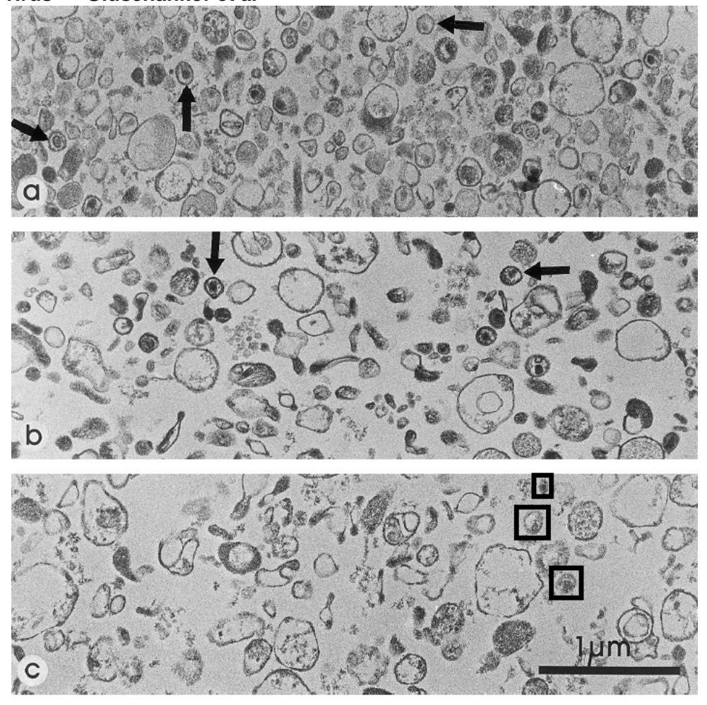
(a) and (b) are purified solutions from an “infected” culture and (c) is a purified solution from an uninfected culture.
The electron micrographs published in the Gluschankof study make it clear that these solutions are in actual fact contaminated to a large degree by cellular debris (bits of the cell culture). Gluschankof et al also cannot avoid admitting this, and that this is the case for both the “infected” and uninfected samples.
For these samples to be called purified retrovirus particle the solutions should contain nothing but virus particles and all particles in the sample should look to be almost exactly the same. This is clearly not the case with these samples.
What is also obvious is that there appears to be virus particles in both the “infected” (marked with arrows) and uninfected sample (marked with squares).
It is also worth noting, that these marked particles are bigger than what retrovirus particles are believed to be and are not the shape they are supposed to be – they lack the cone-shaped cores, lateral bodies and spikes/ nobs protruding from the membrane.
The Gluschankof control demonstrates that “HIV” was never isolated or purified according to the true meaning of the words. It shows that what was claimed to be a solution of pure “retrovirus” particles is in actual fact a soup of particles. This fact in turn, brings into questions the accuracy and legitimacy of all experiments and tests Montagnier and Gallo carried out with these so called “pure” solutions.
The Bess Control Experiment
In 1997, a group from the US National Cancer institute led by Julian Bess, also replicated Montagnier and Gallo’s “purification” procedure and published a paper in which the biochemistry of the “pure HIV particles” ( the 1.16 g/ml band) was analysed. Included in this paper were the results of a control carried out alongside this analysis.
The analysis of the chemistry of the “pure” particles basically amounts to nothing more than determining what the different proteins are which make up the “virus” particles, this is done using a method called electrophoresis.
Electrophoresis is a procedure that is used to separates a mixture of proteins into its individual proteins so that one can determine exactly what proteins make up the mixture. The procedure essentially consists of an electric current attracting the proteins from one side of a gel bath to the other, separating them according to their molecular weights – the lighter proteins moving faster and further along the gel bath, the heavier proteins lagging behind.
Once the proteins have completely separated out from one another the gel is removed and stained. The staining reveals the relative position of each of the separated proteins in the gel and appears as a series of dark, horizontal lines or bands – the protein profile. The thicker and darker the bands the greater the concentration of a particular protein at that position in the gel. One is then able to determine what the particular proteins involved are by comparing the stained results to the stained results of previous electrophoresis experiments carried out with known proteins.

According to Montagnier and Gallo’s work, a solution of “HIV particles” with some cellular contaminates, will show 15 additional proteins to those found in a purified solution of an uninfected cell culture put through the same process. These 15 additional proteins, not found in purified solutions of uninfected cell cultures, are said to be the proteins which constitute the “HIV” particles.
In other words, when comparing the electrophoresis results, one would expect to see 15 horizontal lines in the protein profile of the solution obtained from “infected” culture which do not appear in the protein profile of the solution obtained from uninfected cultures.
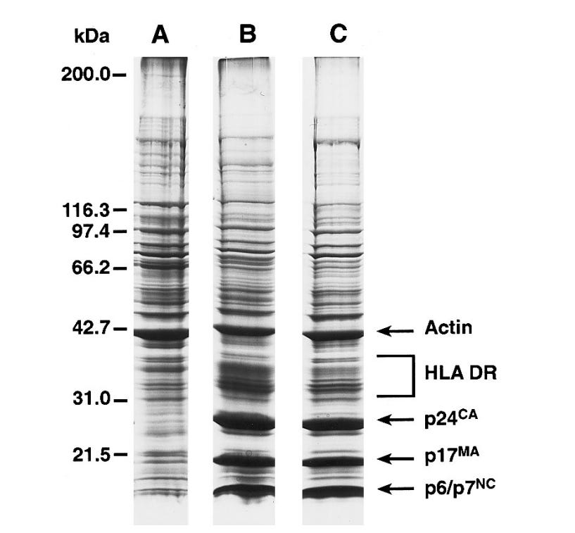
Note, A = uninfected B and C = HIV-infected. Actin and HLA DR = cellular proteins; kDa = molecular weight scale.
Bess carried out electrophoresis on “purified” solutions obtained from three separate cell cultures. “A” in the above image was an uninfected cell culture (the control) and “B” and “C” were cell cultures that had been “infected” with “HIV particles”. As is apparent from the results above, apart from the quantitative (concentration) differences in the results (labelled p6/7, p17 and p24), the protein profiles of B and C are identical to A.
This means that no extra proteins whatsoever, let alone the 15 “HIV proteins”, were found in the solutions obtained from the “infected” cell cultures. The only difference between the samples is that the “infected” samples seemed to have higher concentrations of proteins 6/7, 17 and 24.
Accordingly, the only thing that these results demonstrate is that “infected” cell cultures have greater concentrations of the proteins found in uninfected cultures – i.e more cellular proteins were added not virus proteins.
The Bess control experiment therefore demonstrates that all experiments which made use of the “unique” protein makeup of the “HIV” virus, such as all immunoassay experiments, were fundamentally flawed. The most important being, if there are no unique “HIV” proteins to be found there can be no “HIV antibodies” and thus no HIV antibody tests or HIV genome.

Conclusion
The fact that Montagnier and Gallo did not carry out these simple controls, is not nearly as shocking as the fact that the scientific community was prepared to accept Montagnier and Gallo’s experiments as valid without these controls. This is especially so when one considers the impact the results of these experiments had on the lives of so many people.
The Gluschankof and Bess control experiments demonstrate beyond any doubt how crucial control experiments are for verification of results and how without them virologists (or any scientist really) can claim complete garbage as irrefutable fact.
The fact that the Perth Group picked up on these controls and understood their impact on the accepted science, at a time when no one else in the scientific community seemed to be even the slightest bit sceptical, is a testament to their integrity and the quality of science that they carried out.
Eleni and the Perth Group were truly the OG’s of the no-virus movement and had their work received the attention it deserved at the time of its publication, we might have found the world today to be a totally different place.
Subscribe to dpl
- The above is a simplification of the experiments carried out by the named scientist. For example, all samples obtained from the patients were “isolated” in cell cultures (of different types in some cases) prior to being “purified” by means of density gradient centrifugation but these “isolation” or culturing procedures are not discussed in an effort not to over burden the article. In addition, in some instances, samples underwent more than one round of “purification” and culturing before the ultimate analysis was carried out. All these details and more are set out and discussed at length by Eleni in her manuscript, HIV- A virus like no other, should you wish to review them. The full procedures relating to the control experiments are of course also set out in the Bess and Gluschankof papers linked below.
- It’s interesting to note that Bess et al also published electron micrographs of the “pure HIV” particles “isolated” by means of density gradient centrifugation – see Bess paper for these micrographs. Further, that just as was the case with the Gluschankof electron micrograph, the Bess micrographs also demonstrated that the solution obtained from the 1.16 g/ml bands was anything but pure. However, whereas the Gluschankof micrographs depicted particles of 140 nanometres, the Bess micrographs depicted particles almost double the size measuring almost 240 nanometres. This is problematic as it would mean that Bess’ particles would have a mass 4.7 times greater than the Gluschankof particles, which is more than an unusual finding for one and the same virus. See pg 25-26 and footnote 164 in HIV- A virus like no other.
- E Papadopulos-Eleopulos, 2017. HIV – A virus like no other
- P Gluschankof, 1997. Cell Membrane Vesicles Are a Major Contaminant of Gradient-Enriched Human Immunodeficiency Virus Type-1 Preparations.
- Julian W. Bess Jr, 1997. Microvesicles Are a Source of Contaminating Cellular Proteins Found in Purified HIV-1 Preparations.
- Brent Leung, 2012. The Emperors New Virus? – An Analysis of the Evidence for the Existence of HIV (Documentary)


0 Comments